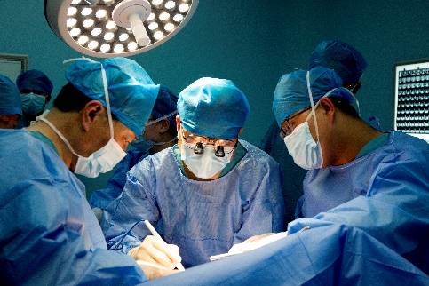
"Artificial intelligence made this 12-hour ex vivo liver resection combined with autologous liver transplantation a great success."
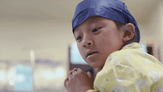
Nearly 80% of her liver was "eaten" by the Echinococcus granulosus parasite. When the 7-year-old girl Zedan was on the verge of death, she traveled thousands of miles to Beijing Tsinghua Changgung Hospital. Two years ago, Zedan was first diagnosed with the disease, and the local hospital announced that it was incurable.

Lu Qian, executive director of the Hepatobiliary and Pancreatic Center, said: "Two veins in Zedan's liver have been eaten by worms, and the human liver has three main hepatic veins: left, middle and right."

The "worms" multiplied in Zedan's liver and formed cysts. Doctors need to avoid these densely packed blood vessels, accurately remove hundreds of parasite cysts, and then rebuild the engulfed blood vessels.
Zedan's entire liver will be removed, and after removing the liver eroded by hydatid cysts, the remaining healthy liver will be re-implanted into the body.
When the liver was exposed during the operation, the true extent of the damage by the tapeworm parasite in the liver was revealed, with countless tiny blood vessels intertwined like a spider web. "We can't see the liver, and the tubes are buried inside," Lu added. In the past, surgeons relied on experience and understanding of organ anatomy to imagine a three-dimensional image in their minds.
This challenge has been a problem for hepatobiliary surgeons for centuries - how to see inside the liver? Artificial intelligence allows hepatobiliary surgeons to "see through" the organ for the first time.
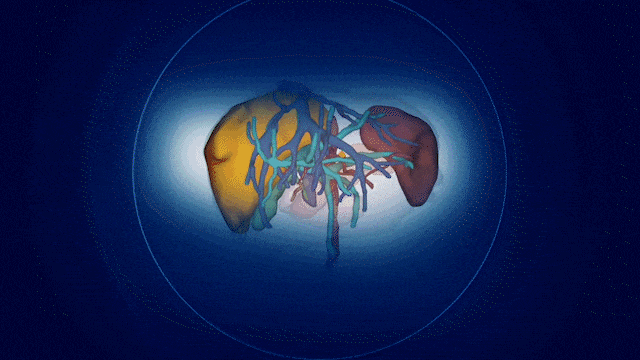
With the software, Zedan's liver was clearly visible, and a three-dimensional image allowed doctors to see through the liver's vascular structure, with different colors representing a group of independent tubes.

The AI system was jointly developed by Dong Jiahong, dean and president of the School of Clinical Medicine, Beijing Tsinghua Changgung Hospital, and Academician You Zheng, to help hepatobiliary surgeons see inside the liver in a virtual environment before surgery, and the three-dimensional image makes the blood vessels clearly visible. "

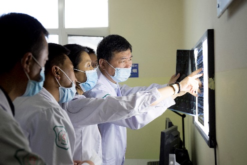
"This digital surgical system accurately restores the patient's liver anatomy and lesions, and reconstructs the past two-dimensional images, such as CT and MRI tomography images, into a three-dimensional computer to form a three-dimensional panoramic structure of the hepatobiliary system." Dong Jiahong said.

Zedan, who traveled a great distance to Beijing to undergo the successful 12-hour surgery has been recovering and has this new technology to thank for a return to her life.
