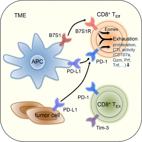
A paper from Chen Dong’s lab entitled “Co-inhibitory molecule B7 superfamily member 1 expressed by tumor-infiltrating myeloid cells induces dysfunction of anti-tumor CD8+ T cells” was published in Immunity on April 3th, 2018. This work revealed that B7S1 expressed by myeloid antigen presenting cells (APCs) initiates exhaustion of CD8+ effector T cells in the tumor microenvironment (TME) and drives T cell exhaustion synergistically with PD-1 co-inhibitory signals. Blockade of B7S1 signaling might improve the efficacy of current anti-PD-1 immunotherapy for cancer.
CD8+ T cells are important effector cells for anti-tumor immune responses. The molecular mechanisms whereby CD8+ T cells become “exhausted” in the TME remain unclear. Programmed Death Ligand-1 (PD-L1) is upregulated on tumor cells and PD-1-PD-L1 blockade has significant efficacy in human tumors; however, most patients do not respond, suggesting additional mechanisms underlying T cell exhaustion.
B7 superfamily member 1 (B7S1), also called B7-H4, B7x or VTCN1, was identified as a co-inhibitory molecule side by Chen Dong, Lieping Chen and James P. Allison’s lab almost at the same time in 2003. Since B7S1 is overexpressed in multiple kinds of cancers, it can be a potential target for cancer immunotherapy. In this study, Dong and his colleagues first detected expression of B7S1 and its receptor in clinical specimens from Hepatocellular Carcinoma (HCC) patients. In the TME, B7S1 was highly expressed on APCs, with it receptor expressed by anti-tumor effector lymphocytes, while PD-L1 was mainly expressed on tumor cells. Moreover, dysfunction of CD8+ T cells correlated with B7S1 (but not PD-L1) expression in HCC tumors.
Next, they investigated the role of B7S1 in anti-tumor immune responses using mouse tumor models. B7S1 inhibition by genetic knockout or antibody blockade suppressed development of mouse HCC (Hepa1-6) and lymphoma (A20 and E.G7). Expression of B7S1 receptor (B7S1R) was mainly observed on CD8+ tumor-infiltrating T lymphocytes (TILs) and B7S1 co-inhibitory signals interfered with cross-presentation between APCs and CD8+ T cells. Then they utilized the E.G7 mouse model to study the mechanism underlying immunosuppression by B7S1. Expression of B7S1R seemed to correlate with infiltration or expansion of CD8+ T cells in the tumor and decreased as tumor progressed at late stage. Interestingly, B7S1R was co-expressed with PD-1 but not Tim-3. Phenotypic and RNA-seq analyses revealed that PD-1+B7S1R+Tim-3-CD8+ TILs were activated, while PD-1+B7S1R-Tim-3+CD8+ TILs were exhausted. Phenotypic analysis of E.G7-bearing wildtype vs. B7S1 knockout mice showed that B7S1 inhibition could significantly enhance expansion and effector functions of CD8+ TILs. What’s more, B7S1 co-inhibitory signals drove T cell exhaustion through transcription factor Eomes as revealed by in vitro and in vivo functional assays.
Since B7S1R and PD-1 were co-expressed on CD8+ TILs, and B7S1 inhibition led to compensatory up-regulation of PD-1, they speculated that B7S1 and PD-1 signaling might drive T cell exhaustion cooperatively and co-blockade of these two pathways might further enhance tumor immunity. Therefore, they compared the efficacy of anti-B7S1 or anti-PD-1 monotherapy with combinational therapy in E.G7 and Hepa1-6 mouse model. As a result, anti-B7S1 and anti-PD-1 combinational therapy inhibited tumor growth more potently. In order to understand the mechanism underlying the synergic effects, they compared open chromatin regions and transcriptomes of OVA-specific CD8+ TILs from E.G7-bearing mice treated with control, anti-B7S1, anti-PD-1 and anti-B7S1+anti-PD-1 antibodies by ATAC-seq and RNA-seq analyses. Majority of epigenetic regulations, gene expressions and signaling pathways were co-regulated by B7S1R and PD-1. B7S1R and PD-1 co-expressed on CD8+ TILs might act on shared downstream components to suppress their anti-tumor activity coordinately. Therefore, combinational blockade of B7S1 and PD-1 might improve the clinical efficacy of the current anti-PD-1 therapy for cancer.

B7S1 and PD-1 signaling drives T cell exhaustion cooperatively
PD-1 and B7S1R co-expressed on CD8+ TILs might receive inhibitory signals from tumor cells or APCs, and induce T cell exhaustion via acting on shared downstream components.
Dr. Chen Dong is the corresponding author and Ph.D. candidate Jing Li is the first author of this paper. Researchers from Tsinghua University, MD Anderson Cancer Center, University of Texas, University of Toronto and China-Japan Friendship Hospital also contributed to this study. This study was funded by Beijing Municipal Science and Technology and National Natural Science Foundation of China (NSFC).
Link: http://www.cell.com/immunity/fulltext/S1074-7613(18)30115-8
